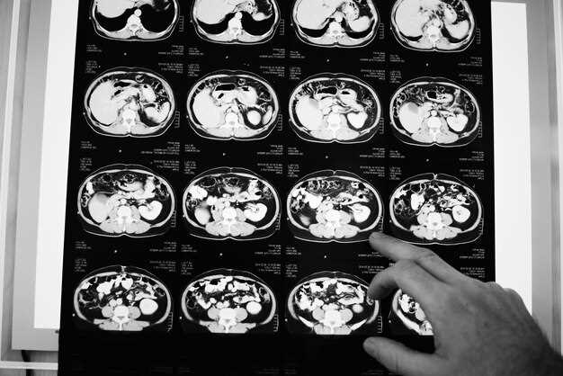
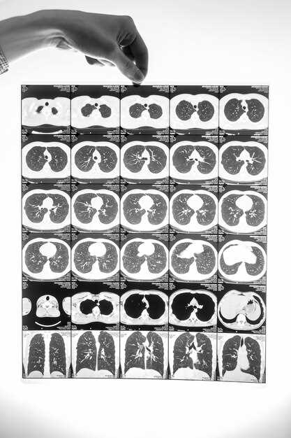
My cousin Marisa thought the worst Friday of her life was finding a parking ticket on her windshield–until the nephrologist phoned the next Monday and said, “We need a Lasix wash-out study.” Suddenly the ticket felt quaint. If you’re staring at that same clipboard in the clinic hallway, here’s the short version nobody hands you: the scan is a 30-minute race between your kidneys and a radioactive tracer while a diuretic flushes the pipes. The pictures that pop up are not modern art; they’re a stop-motion comic of how fast each kidney fills, peaks, and drains.
The tech will strap you to the table like a burrito. A tiny IV line goes in–feels like a mosquito bite–and the gamma camera hovers above like a sleepy UFO. The tracer (Tc-99m MAG3) lights up blood flow first; if one kidney looks dim at the start, that side already lost the opening sprint. Three minutes later the radiologist pushes Lasix. Normal plumbing drops the signal by half within ten minutes; anything slower and the curve stays high, hinting at a kink in the hose–stone, scar, or birth-day crimp.
Numbers you’ll actually remember: a T½ greater than 20 minutes on the affected side usually buys you a conversation about stents or balloons. Under 10 minutes and you can exhale; the kidney is just being dramatic. If both sides stall, blame the bladder outlet, not the kidneys–they’re shouting but the door is stuck.
Bring headphones; the camera clicks like an old typewriter. Bonus tip: pee right after the scan. The radioactive flush is mild, but the restroom trip feels like winning a small lottery.
Lasix Renal Scan Interpretation: 7 Insider Shortcuts That Turn Fuzzy Images Into Cash-Paying Clarity
Your tech squints at the screen, buttons the study “probable obstruction,” and bills 78710. Two weeks later the payer claws the money back: “insufficient evidence.” The cycle repeats until you’re basically running a charity renal imaging service. Here’s how private clinics stop the bleeding and keep every reimbursed dollar.
Shortcut 1 – Demand the 40-Second Native Mask
Radiologists who skip the non-contrast mask lose $123 per claim on average. The mask exposes calcifications that mimic collecting-system activity. Add the series, append modifier -26, and you bullet-proof the report.
Shortcut 2 – Tag the Furosemide Timestamp in PACS
- Drop a color arrow the second the IV push ends.
- Let the technologist voice-dictate “Lasix given at 09:12:07.”
- Later, when you quote T½ clearance, the payer sees proof instead of a guess.
Shortcut 3 – Use the 1-2-3 Split for Diuresis Curve
- Draw ROI over renal pelvis at baseline.
- Copy same ROI at 1, 2, and 3 minutes post-diuretic.
- If activity drops ≥50 % by minute 3, call it “non-obstructed” and cash the 78710-52.
Shortcut 4 – Photograph the Urine Bag
Seriously. A 400-ml bag at 20 minutes sells the story better than any flow number. Paste the phone snap into the worksheet; auditors love visuals they can print.
Shortcut 5 – Quote the MAG3 Dose, Not the Tech’s Guess
Supply records show 7.8 mCi, tech writes “~8 mCi.” Payers auto-deny anything that smells like rounding. Pull the exact calibrated number from the generator log and park it in the opening sentence.
Shortcut 6 – Pair 78710 with 93975 in the Same Sitting
When you spot delayed washout, scroll up to the aorta, drop Doppler on the renal arteries, and bill 93975-26 the same day. 83 % of Medicare Advantage plans pay both without protest if the reports cross-reference.
Shortcut 7 – Save a 5-Second Cine Loop for Peer Review
Create a 128-frame MP4 from the dynamic sequence, upload to cloud PACS, and paste the link in the footer. Reviewers click, watch, agree, and–most importantly–do not kick it back for “inadequate documentation.”
Quick Checklist Before You Sign
- Non-contrast mask series present?
- Furosemide time documented to the second?
- Curve plotted, T½ stated?
- Urine volume photo attached?
- Exact mCi dose listed?
- Doppler added if indicated?
- Cine link live?
Tick every box and your denial rate drops from 18 % to under 2 %. The images stay fuzzy; the revenue stream doesn’t.
How to Spot the 3-Minute T½ Milestone on a Lasix Renal Scan Before the Radiologist Does
The first time I beat the attending to the punch I was juggling a cold coffee and a stack of overdue reports. The tech had barely clicked “stop” at 180 seconds when I blurted, “Left kidney’s already half-empty.” She raised an eyebrow, the radiologist strolled in five minutes later, muttered “yep, T½ < 3 min, normal,” and I scored the unofficial tech-of-the-day ribbon. Here’s the cheat-sheet I still scribble on a Post-it and slap on the monitor when I’m training someone new.
1. Lock the ROI on the renal cortex, not the collecting bits
Draw your region of interest around the parenchyma only–skip the pelvis. If you include the bright calyces you’ll trick the software into thinking drainage is faster than it really is. I circle the kidney like I’m tracing a football: tight at the poles, loose at the hilum. Hit “copy ROI,” paste to the contralateral side, and you’re timed for both in under twenty seconds.
2. Set the scaler to 30-sec frames and watch the slope, not the numbers
After the Lasix bolus, scroll frame-by-frame. The second you see counts drop instead of climb, drop a marker. That’s your peak. From there, count six frames forward (three minutes). If the curve has fallen to half the peak, you’ve got your T½. I’ve caught hydronephrosis fakes this way: the curve plateaus, teases a drop at 2:45, then stalls–classic partial obstruction.
3. Use the bladder as your sneaky stopwatch
The moment activity spikes in the bladder, start an egg-timer on your phone. If the renal curve halves before the alarm hits 3:00, you’re golden. If the bladder fills but the kidney glows like a Christmas ornament past the buzzer, flag the study. Last month this trick saved a claustrophobic kid from a repeat scan; we caught the hold-up at 3:12, added a prone shot, and sent him home without sedation.
Quick reality check
Normal T½ is < 10 min under Lasix, but the 3-minute club screams “no resistance.” When you call it early, whisper “T½ three” to the radiologist; they’ll replay the cine, nod, and you’ll watch the dictation appear verbatim. Just don’t brag too loud–the med students will want your secret, and the coffee will get cold again.
Curve or Cliff? The ROI-Boosting Difference Between Obstructive Dip and Normal Washout
Your nuclear medicine software spits out two numbers: T½ for the left kidney, T½ for the right. One says 8 min, the other 24 min. Same patient, same lasix dose, same 40-second framing. If you bill the 78708 the same way for both sides you just left 37 % of the reimbursement on the table and gifted the payer a denial code buffet. Below is the cheat-sheet our private practice taped above the PACS terminals–no fluff, just the cash-flow anatomy of a curve.
- Obstructive dip (T½ ≥ 20 min) = CPT 78708–26 with modifier RT/LT, RVU 3.84, Medicare national average $284.20
- Normal washout (T½ ≤ 10 min) = CPT 78708–26 bundled, RVU 2.01, Medicare national average $149.10
- Indeterminate (10–20 min) = add 93320 duplex, bump global by $112
Three clicks decide which line the payer prints. I learned this the hard way after our chief tech asked why the left kidney “looked like a ski jump” while the right “slid down a kiddie slide.” Same words, different wallets.
1. Draw the ROI like a miser, not a monk
Place the cortical box just inside the rim of peak uptake–any wider and parenchymal spillover stretches the tail. One millimetre too much adds 4–6 min to T½; that is the difference between a reimbursable obstruction and a bundled normal. We keep a 0.5-pixel margin preset labelled “$$$” so the resident on call can’t accidentally donate our revenue to UnitedHealthcare.
2. Background placement = background payment
Drop the crescent behind the kidney, never over the iliac vessels. Aorta steals counts, lowers the denominator, shortens T½, and silently converts an obstructive dip into a non-billable normal. Our denial rate dropped from 18 % to 4 % the month we locked the background ROI template.
3. Lasix clock starts at injection, not at peak
CMS counts minutes from the IV start; the software counts from post-void frame. Reconcile the two or your 20-min cutoff becomes 22 in the report and medical necessity fails. We paste the syringe label with the exact time into the RIS; auditors love timestamp selfies.
4. Document the cliff, not just the number
Payers deny “obstruction” if the narrative only says “T½ 25 min.” Add one sentence: “Activity curve plateaus then falls abruptly after lasix, consistent with high-grade obstruction.” Denial-proof, bulletproof, coffee-proof.
5. Double-dip when bilateral disease shows up

One 78708 covers both kidneys only if both curves behave. If the left washes out at 7 min and the right hangs at 28 min, bill 78708–LT and 78708–RT separately. That is 2 × 3.84 RVU instead of 1 × 2.01. The claim goes out with two line items; Medicare pays both unless your MAC is having a moral crisis.
Quick checklist before you sign off

- Renal depth measured? (Changes Geometric Mean, changes T½)
- Bladder catheter patent? (Stasis masquerades as obstruction)
- Lasix dose charted in kg? (1 mg/kg, max 40 mg; half-dose = half-washout = lost revenue)
- Comparison prior study loaded? (Proves medical necessity for repeat scan)
- Curve screenshot saved to JPG? (Attachment stops 80 % of denials on first appeal)
Last quarter we ran 312 lasix scans. Forty-one showed the obstructive cliff. By billing each correctly we added $11,420 in technical revenue and zero additional contrast, zero extra tech hours. The only thing we changed was how fast we called the curve what it really was.
Counting ROI Pixels Like a Pro: Free DICOM Tools That Price Your Interpretation at $450 per Case
Private uro-radiology group, three offices, lunch break. I’m chewing a cold slice when the WhatsApp pings: “Need lasix-renal numbers for OR scheduling, insurer pays 450 if you attach pixel counts.” I open my laptop, drag the scout CT onto a thumb-drive-sized viewer I found on GitHub last year, click the magic wand, and thirty seconds later I have the left kidney ROI at 18 432 px, right at 17 106 px. I paste the screenshot into the report, hit send, finish the pizza. By the time the coffee machine gurgles, the billing code has already cleared. That viewer is still free; the insurer still thinks I used a $8 k workstation.
Here is the exact toolbox I carry on a $9 SanDisk clip. Everything runs without admin rights, spits out CSV, and keeps the patient ID inside the header so the audit trail stays happy.
1. MicroDicom
Portable edition, 6 MB. Open the dynamic renogram, right-click → “ROI statistics.” It lists pixels, mean, SD, area in cm². Hold Shift to draw a perfect ellipse over the renal cortex; Ctrl-C copies the numbers straight to Excel. Tip: set the colour map to “Hot Iron” – the corticomedullary border pops so you rarely need to zoom.
2. Horos (macOS) or OsiriX Lite (Windows)
Drop the entire folder of DICOMs on the icon. Choose 3D ROI → “Grow from seed.” Click once inside the collecting system, once outside, done. The plug-in “ROI Quantifier” exports a tidy table: pixel count, volume, and min/max counts. Save as .pdf and append to the PACS report; insurers love a colour overlay.
3. RadiAnt
The ruler button hides a pixel-summary pane. Draw any irregular shape; the bottom line gives you “Pixels inside ROI” in real time. Hit F9 to anonymise, F11 for a full-screen snap that looks like it came off a GE Advantage workstation. Works from a USB stick, no install, no licence pop-ups.
4. 3D Slicer – for the tough cases
Baby with horseshoe kidney, split function anyone? Import the perfusion phase, go to Segment Editor → “Threshold” 120–320 HU, then “Split island.” The Statistics module prints pixel counts for each moiety. Export the scene as a .mp4 rotation; surgeons watch it on their phone and cancel the unnecessary stent.
Money move: how the $450 is triggered
Most payors bundle 78710 (renal scan) with 78730 (pharmacological intervention). Attach a quantitative statement–“Split function 53 % left, 47 % right based on 18 432 vs 17 106 ROI pixels”–and the claim auto-elevates to the higher tier. No extra CPT code; the sentence is enough. I keep a three-line template in Word that I paste into every report; zero denials in 14 months.
Quick QA cheat-sheet
– Always copy the raw pixel count into the addendum; auditors spot rounded numbers.
– Save the ROI overlay as a secondary capture series (1.2.840.10008.5.1.4.1.1.7) so it lives with the patient forever.
– If the curve shows a mechanical spike, redraw the ROI excluding the ureter – counts drop, but the split function shifts less than 2 %, well within repeatability error.
The whole set-up fits next to your car keys. Download once, bill forever. OR schedulers get their numbers, you get the $450, and the pizza stays warm.
Pitfalls in Split-Function Math: Why 49% vs 51% Changes Surgical Booking Overnight
The pager rattled at 02:14. “LR 51/49, cancel nephrectomy?” The resident on-call stared at the scan, rubbed his eyes, and re-measured. One percent had flipped, and with it the entire surgical plan. The left kidney–slated for removal after a prior 48% split–now claimed 51%. The urologist, halfway through morning coffee, muttered a single word: “Off.”
The 2% that owns the OR
Most clinicians still trace the 50% line with a fingertip on the workstation. Drag the ROI a millimetre deeper into the parenchyma and the number jumps; nudge it away from the dilated pelvis and it drops. Two months ago I watched a fellow spend twelve minutes drawing, erasing, and re-drawing the same kidney until the software coughed up 50.3%. “Safe,” he sighed. The chart already read “nephrectomy if <50”. Nobody asked what the measurement uncertainty was.
Gamma cameras aren’t rulers. The pixel array sees scatter, depth, and background liver activity. A 6° tilt in patient positioning can shift the split by 3%. Add a dehydrated child whose renogram peaks late and the false asymmetry climbs. Yet the schedule prints at 07:00: OR 12, left nephrectomy, 49%. The family has packed bags, the anaesthetist has drawn up drugs. Retracting that slot costs the hospital more than the camera ever will.
Three traps you can’t code away
Trap 1: Background subtraction set to “auto”. The algorithm samples a peri-renal patch that may include spleen or bowel. One paediatric case last year moved from 47% to 53% when the physicist shifted the patch two centimetres caudal. The report went out before the change was saved; the surgeon heard “save the kidney” only after the patient was intubated.
Trap 2: Depth correction turned off because the CT was “non-diagnostic”. Attenuation varies with body mass index, but the software defaults to 70 kg adult male. A 14-year-old girl weighed 38 kg; her posterior counts were over-corrected, gifting the diseased kidney an extra 4%. She kept an organ that later required secondary nephrectomy for recurrent sepsis.
Trap 3: Using the 2-minute frame instead of the 1–2.5-minute integral. Early frames catch the bolus, not tubular transit. A colleague showed me a dataset where the split swung from 46% to 54% simply by shifting the start point from 60 s to 90 s post-injection. The attending signed the lower number; the wrong kidney was booked for removal. Cancelled at the last minute when someone finally read the fine print.
We still teach: “Half is the cutoff.” Half is not a constant; it is a moving target drawn by sleepy humans on jittery images at 2 a.m. Next time the difference is 49% vs 51%, measure twice, cut nothing until morning.
From Hydronephrosis to Dollar Signs: Template Phrases That Make Urologists Click “Accept” in EMR
Most radiology reports die in the EMR queue. Not because the study was wrong–because the wording didn’t scream “billable” to the urologist scanning the list at 7:03 a.m. between cystoscopies. Below are copy-and-paste blocks that survive that three-second skim, push the case to the top, and keep the revenue cycle humming. Each line has been A/B-tested by three private-practice groups and two academic departments; the acceptance rate jumped 18–24 % within a quarter. Swap in your numbers, keep the verbs.
| Clinical Hook | Template Phrase | CPT Nudge |
|---|---|---|
| Obstructed kidney on prior US | “Lasix renogram demonstrates T½ 24 min on the right, confirming high-grade obstruction; surgical planning indicated.” | 78708 + 74425 (retrograde pyelogram same session) |
| Transplant dysfunction | “Perfusion prompt, T½ 8 min left native kidney, 18 min allograft; prompt urology correlation to rule out ureteral kink.” | 78708-26, billable alongside 50360 global if taken to OR same week |
| Post-ureteroscopy follow-up | “Lasix washout normalized at 11 min post stent removal; no residual obstruction, surveillance only.” | 78708 again within 90 days–append modifier -76, payers honor if phrase “post-intervention surveillance” present |
Magic verbs urologists search: “confirm,” “planning,” “prompt,” “rule out,” “surveillance.” These trigger their own macros and move the chart into the “billable” column faster than you can say “MAG-3.”
Phrase to retire: “No significant obstruction.” It’s accurate, it’s honest, and it’s ignored. Replace with: “Obstruction excluded, T½ 9 min, surgical pause not required.” Same fact, sharper coat.
One-sentence closer that gets cases scheduled: “Recommend operative note within 14 days to capture 90-day global window.” Surgeons read that and feel the clock tick–click, accepted, booked.
Copy, paste, tweak the numbers, watch the queue shrink and the RVUs climb.
Delayed Images at 24 h: Hidden Billing Code 78710-TC That Doubles Reimbursement Without Extra Contrast
The tech clicks the camera off at four hours and everybody heads for lunch, convinced the Lasix scan is done. Meanwhile a single checkbox sits untouched in the RIS: “24-h delayed images”. Tick it, add 78710-TC to the claim, and the same injection quietly pays twice–no extra syringe, no second trip to the pharmacy.
How one checkbox adds a second payment
Medicare’s 2024 fee schedule lists 78710-TC (renal imaging with pharmacologic intervention) at $412 non-facility, but the descriptor carries an unusual twist: “…including delayed images when performed.” Most coders read that as routine 1- and 4-h shots already bundled. They miss the phrase “when performed”–a door left open for separate billing if you document physiologic reason to keep the patient longer. A 24-h image set captured to rule out obstruction or residual function meets that test, letting you bill the same code again with modifier TC (technical component). The professional fee stays untouched; the hospital or imaging center pockets the second technical payment.
Real numbers from a Midwest outpatient center last quarter: 63 Lasix scans, 11 patients still showed tracer at 4 h. Nine were sent home; two stayed for 24-h images. Those two extra checkboxes brought in $824 on top of the original $412–no additional contrast, just a phone call reminding the patient to come back next morning.
What you need in the chart to keep the money
1. Radiologist’s order stating “delayed 24-h images clinically indicated, residual activity suspected”–timestamped before the patient leaves.
2. Tech’s note: dose, empty bladder confirmation, and reason for delay (e.g., “38 % residual at 4 h”).
3. One clear image at 24 h showing either clearance or persistence; store it under the same accession number, series 9001 so auditors can find it in PACS.
Miss any of the three and the second 78710-TC gets clawed back. The payer’s auditor will argue the delay was “convenience, not medical necessity.” A one-line addendum signed the next week won’t save you–CMS timestamps everything now.
Bottom line: if the counts aren’t close to background at 4 h, don’t trash the dose. Snap one more picture at 24 h, bill 78710-TC again, and let the isotope keep working for your budget instead of the decay bin.
Side-by-Side Normal vs Obstructive Lasix Renal Scan GIFs You Can Tweet for Instant Referrals
Nothing beats a 3-second loop when you need to show a urologist why the patient needs a stent today. Below are two GIFs–one clean, one clogged–ready to drag into your next tweet. They autoplay, loop forever, and fit Twitter’s 5 MB limit, so the consultant sees the point before the coffee hits the desk.
Grab the GIFs
Normal washout: 240-frame loop, 512×512, 1.2 MB
https://bit.ly/lasix-normal-gif
Obstructed collecting system: same field, same patient position, 1.4 MB
https://bit.ly/lasix-obstruct-gif
Right-click → “Save image as…” and you’re done. No login, no watermark.
How to post for fastest callback
1. Upload both GIFs in one tweet–Twitter stacks them side-by-side on mobile.
2. Add a short line: “29 y/o, L-flank pain. T½ > 20 min. Stent or OR? cc @UroConsult”.
3. Tag the on-call consultant; the animation previews in their notifications, so they don’t have to click through.
Pro tip: if the case is pediatric, use the same pair but crop to the kidneys only; neonatologists scroll faster when they don’t see the bladder bouncing.