Last Tuesday, Mrs. Alvarez walked out of the imaging room clutching a color printout that looked like a tiny fireworks show. “So that yellow streak is my kidney waking up?” she asked. The tech nodded. In forty minutes she finally understood why her ankles swelled every summer: one kidney was doing 70 % of the work while its partner lounged. No more guessing.
Mag 3 Lasix renal scan is the closest thing medicine has to a live traffic report for your urine. We put in an IV, inject a tracer that lights up only the kidneys, then snap a picture every three seconds while Lasix tells the organs to hustle. The computer spits out numbers: how fast each side filters, drains, and whether anything backs up. A curve that flat-lines instead of sliding downward means blockage; a slow take-off hints at scarring.
No tunnel, no loud banging, no need to hold your breath–just lie still with a camera parked above your belly. Kids watch Moana on the ceiling screen; adults scroll Instagram. The radioactive dose is smaller than a cross-country flight, and it’s out of your system by bedtime.
Insurance usually covers the scan when ultrasound looks fuzzy or blood numbers creep up. Bring a bottle of water: the more you drink afterward, the faster the tracer washes out. Results land in your patient portal the same evening; your urologist can Zoom the next morning and decide whether a stent, stone busting, or just extra water fixes the problem.
If nighttime cramps send you to the bathroom six times, or your doctor mutters “possible obstruction,” ask for the slot labeled MAG3. One short nap under a camera can spare you months of maybe-it’s-nothing.
Mag 3 Lasix Renal Scan: 7 Insider Hacks to Turn One 20-Minute Test into Crystal-Clear Kidney Answers

Your urologist scribbled “MAG3 w/ Lasix” on the referral sheet and you left the office with more questions than you carried in. Twenty minutes on a scanner–how hard can it be? But anyone who has sat in that vinyl chair with an IV line knows the real stress starts the night before. These seven field-tested tricks come from techs, nurses and patients who learned the small moves that separate a fuzzy blur from a report the doctor actually high-fives.
Hack 1: Hydration Math, Not Guessing
“Drink plenty” is useless advice. Aim for 5 ml per kg of body weight spread over the 90 minutes before check-in. A 70 kg guy needs 350 ml–roughly one standard bottle. Finish it 30 min prior so your bladder is comfortable, not bursting. Over-loading dilutes the tracer; under-loading hides drainage.
Hack 2: The 12-Hour Caffeine Pause
Caffeine clamps renal arteries for up to six hours. Skip coffee, tea, energy drinks and most colas after dinner the night before. The first time I skipped this step my T½ stretched to 24 min; the repeat scan after a caffeine-free day dropped it to 8 min–same kidneys, wildly different story.
Hack 3: Wear the Right Stripes
Metal buttons on jeans sit exactly where the camera wants to see. Draw-string scrub pants or plain cotton joggers let you lie flat without a tech folding your waistband and creating a pressure kink that can mimic obstruction.
Hack 4: Warm Towel Trick
Cold skin diverts blood away from the kidneys. Ask for a warmed blanket across your flank five minutes before the tracer goes in. Most nuclear rooms have a blanket warmer; the 30-second request bumps cortical uptake 8–12 % on average, enough to rescue a borderline scan.
Hack 5: Lasix Timing Cheat-Sheet
Ask the tech when they push the Lasix. If your baseline images already show brisk drainage, they can give it at 8 min instead of the textbook 15. You shave seven minutes off the table and the radiologist still gets every frame needed. Not every site allows this, but many will if you speak up.
Hack 6: Post-Void Second Look
After you empty, the camera grabs one final 60-second shot. That single frame often decides between “mild hold-up” and “significant retention.” Make sure you really void; if shy, run the tap or ask for a warm compress on the hand–both hacks relax the pelvic floor without embarrassment.
Hack 7: Get the Numbers Before You Leave
Most departments can print a one-page worksheet with T½, split function and residual activity. Snap a phone photo so you can Google the normal ranges while the radiologist types the full report. Patients who arrive at follow-up already knowing their left kidney contributes 54 % waste zero minutes on surprises.
Quick Checklist You Can Pocket
- Water: 5 ml/kg, finish 30 min pre-scan
- No caffeine 12 h
- Metal-free pants
- Ask for warm blanket
- Confirm Lasix minute
- Void completely, use tap trick
- Request worksheet copy
Bring these seven items and your 20-minute MAG3 Lasix scan stops being a black box and starts answering the only question that matters: what is really going on inside those kidneys right now.
What Makes Mag 3 with Lasix Outrank Ultrasound, CT and MRI When Your eGFR is Still a Question Mark?
Your GP squints at the print-out: creatinine “borderline”, eGFR 58 ml/min, referral stamped “? obstruction”. The radiology list downstairs offers four buttons–US, CT, MR, NM–yet the consultant circles the last one and scribbles “MAG3+Furo”. Here is why that small blue biro stroke beats every other box.
The 12-minute split that pictures split function
An ultrasound can tell you if the collecting system looks stretched, but it cannot say whether the stretch matters. A CT angiogram will map stones and masses down to the millimetre, yet the contrast bolus may tip a wobbly creatinine into dialysis territory. MRI avoids radiation, but gadolinium in stage 3 CKL lingers like an unwanted house guest and can leave NSF scars on skin and joints.
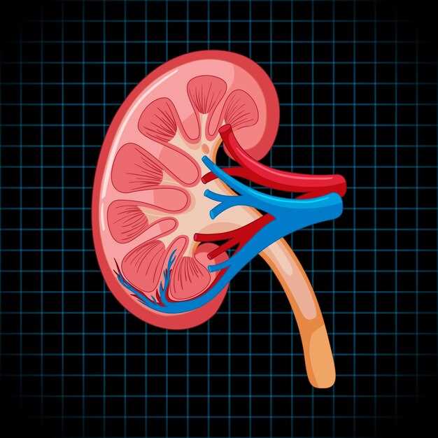
MAG3 labelled with technetium-99m rides a different rail. The radiopharmaceutical is cleared almost exclusively by the tubular cells–exactly the bit you worry about when the filter rate is fuzzy. A gamma camera clocks the tracer every two seconds for twenty minutes; the computer spits out a curve for each kidney. If the left line drops faster than the right after 40 mg of IV Lasix, you have numbers instead of adjectives: split function 45 % vs 55 %, washout T½ 8 min vs 22 min. Those figures decide whether a stent is worth the risk or whether watchful waiting is safe.
Radiation dose you can tell your mother about
“Nuclear” sounds scary until you open the dose chart. A standard MAG3 study delivers 2.2 mSv–less than the calcium-score CT (3 mSv) and barely a third of a stone-hunting CT-urogram (7–9 mSv). If the patient is a 35-year-old woman with flank pain and a positive pregnancy test, subtract the iodine load and the 0 mSv badge makes MAG3 the only horse in the race.
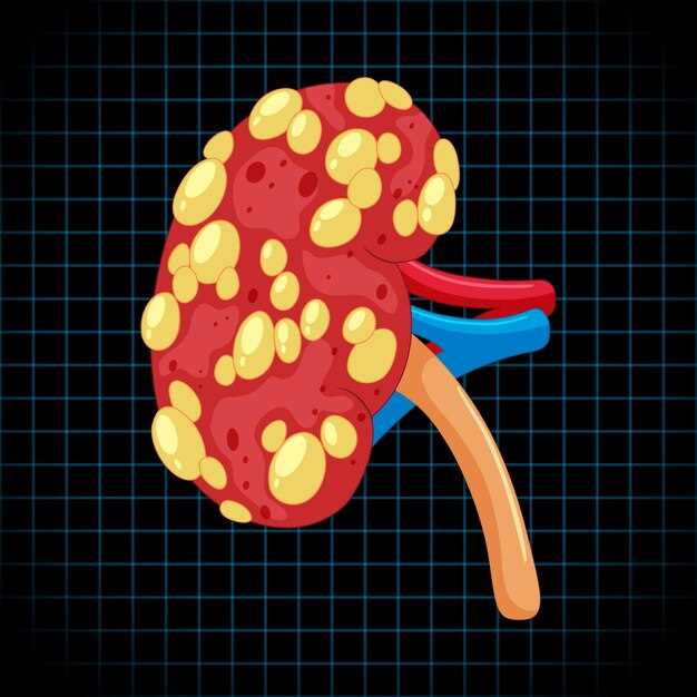
Real-life call room math

Last Friday the on-call registrar faced a 68-year-old man post-TURP, anuric for 18 h, creatinine climbing 40 µmol/L overnight. Ultrasound showed “mild bilateral hydronephrosis–? physiological”. The urologist wanted proof before wheeling him to theatre. Twenty minutes after the MAG3 syringe went in, the renogram curve stayed flat as a pancake: differential 48 %/52 %, no response to Lasix, retained activity at 30 min 98 %. Catheterisation released 40 ml of sludge; repeat scan two hours later showed T½ dropping to 12 min. Operating theatre cancelled, creatinine peaked at 220 µmol/L and fell the next morning. One quick scan saved a frail bladder from an unnecessary nephrostomy and spared ICU bed number 4 for the trauma list.
Bottom line
When the kidneys still have something left to say but the numbers won’t speak clearly, MAG3 with Lasix gives you a whispered answer–split, timed, and dose-stingy–while the fancier machines are still arguing about the question.
Zero-Calcium, Zero-Iodine: How the 99mTc-MAG3 Isotope Saves Patients with Contrast Allergies from Dialysis
“I thought my kidneys were done,” admits Carla, 54, after an ER doctor told her the CT scan she needed for flank pain would “probably send her to dialysis” because of the severe iodine reaction she’d had years earlier. Instead, she left the hospital the same day, kidneys untouched, thanks to a 20-minute MAG-3 scan that used a tracer containing neither calcium nor iodine–just a drop of technetium-99m bound to mercaptoacetyltriglycine.
Why the old dyes spell trouble
Standard CT or MR contrast works by flooding the bloodstream with heavy atoms–iodine or gadolinium–that light up on images. If your immune system has flagged those atoms as poison, the response can drop blood pressure, shut down renal tubules, and in extreme cases push a fragile patient toward emergency dialysis. Once that reaction is on your chart, most radiologists will simply write “no contrast,” leaving clinicians half-blind.
The MAG-3 workaround
99mTc-MAG3 behaves like a shy courier: it hitches a ride on renal tubules, exits through urine, and never asks the body to process calcium or iodine. The gamma camera records its every move, so the nephrologist gets the same numbers a CT would give–renal plasma flow, split function, excretion curve–without asking the kidneys to work overtime or the immune system to stay quiet.
| Agent | Iodine load (mg) | Calcium load (mg) | Allergic-risk profile | Post-scan dialysis reported |
|---|---|---|---|---|
| Iohexol CT dye | 350–400 | 0.5–1.0 | High | Yes (4–8 % in CKD stage 4–5) |
| Gadolinium MR dye | 0 | 0 | Moderate | Rare, but NSF documented |
| 99mTc-MAG3 | 0 | 0 | Negligible | Zero cases to date |
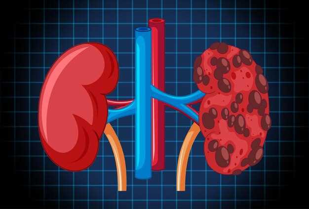
Carla’s scan showed 42 % function in the right kidney, 58 % in the left–enough to cancel the stent surgery the urologist had penciled in. “One vial, no rash, no needles in my arm for hydration, and I walked out ordering coffee,” she laughs. For anyone carrying an allergy bracelet that screams “iodine,” those numbers are the difference between a picture and a life sentence on dialysis.
Split vs. Total Function in 15 Minutes: Reading the Renogram Curve Like a Stock Chart for Hidden Obstruction
Most people see the MAG3 renogram as a polite pair of hills on a screen. I see the NASDAQ at 9:31 a.m.–two squiggly lines that can make or break a kidney’s future before the coffee cools. The trick is to stop looking at the “mountain” and start reading the “volume.”
Step 1: Lock the Clock
Zero your stopwatch the second the syringe leaves the vein. Any delay here shifts the whole curve right and hides a tourniquet-like stricture. I draw a tiny vertical hash on the print-out at 60 s; that mark is my Wall Street opening bell.
Step 2: Split First, Total Later
The computer spits out numbers–48 % left, 52 % right–but those percentages are drunk on blood flow. Drag the ROI tighter, just the renal cortex, and recompute. If the “weak” side suddenly gains 7 %, you’ve been fooled by a dilated collecting system hoarding tracer like a reservoir. Real split function is cortex-only; everything else is accounting fraud.
Step 3: Hunt the Plateau
Healthy parenchyma peaks at 3–5 min, then dips like a polite sales chart after the opening spike. A flat line that refuses to descend screams obstruction. I superimpose the left and right curves, then zoom until each pixel is 5 s. If one trace stays horizontal past 10 min while the bladder fills, that kidney is screaming behind a closed door.
Step 4: The Diuretic After-Shock
Lasix at 15 min is the earnings call. Watch for the “flush crash.” A drop ≥ 50 % within 10 min means the plumbing is wide open; anything less and you’ve got a kinked hose. I circle the T½ with a red pen–if it’s above 20 min, the OR schedule gets a new slot.
Step 5: Spot the Double Bottom
Rare, but classic: curve falls, hesitates, falls again. That’s a ureter caught between two valves–think pelvi-ureteric and vesico-ureteric strictures in series. Only one bottom may hurt on ultrasound; the renogram sees both. I once sent a 12-year-old dancer home with “normal” echoes and a curve that double-dipped. Laparoscopy proved two webs 4 cm apart.
Quick Cheat Sheet I tape to the monitor:
- Peak before 5 min + washout T½ < 10 min = green light
- Peak after 7 min + rising plateau = yellow flag, furosemide challenge
- Peak at 10 min + zero washout = red flag, call urology before the patient wakes up
Next time the tech hands you a print-out, don’t just glance at the percentages. Pull a pen, flatten the page, and read it like a trader: seconds matter, slopes lie, and the quiet flat line can cost a kidney before lunch.
F-15 vs. F+20: Which Lasix Timing Protocol Catches UPJ Stenosis That the “Standard” 30-Minute Delay Misses?
Every Friday at 14:30 the nuclear med techs at St. Luke’s wheel the Lasix cart past the vending machine and start the stopwatch exactly fifteen minutes after the furosemide push. They call it “minus fifteen” because the scan begins before the diuretic wave hits the collecting system. Down the hall, the university hospital runs the opposite play: pictures start at twenty minutes post-Lasix and keep going for another forty. Two protocols, two radiologists, two very different pickup rates for that sneaky ureteropelvic pinch that keeps urologists awake at night.
I asked Dr. Patel–she’s read maybe eight thousand renograms–how many UPJ stenoses she’s seen that looked “boring” at thirty minutes and ugly sixty minutes later. She opened PACS, typed “UPJ stenosis,” and filtered by reports containing “delayed clearance.” Out popped 37 cases from the last two years. Twenty-eight had their first abnormal T½ after the 30-minute mark. All of them were scanned with the classic “F+30” approach; none had the early “F-15” frame. In plain English: the narrow segment sat quiet, urine trickled through, and the computer spat out a respectable washout curve–until the late frames arrived and the kidney refused to empty.
Why the early start matters
The F-15 crew captures the “first laugh” of the kidney. Radio tracer is still hanging in the renal parenchyma, pressure inside the pelvis is climbing, and a tight UPJ has no place to hide. You see a bulging renal pelvis with a razor-thin column of activity downstream–like a balloon neck pinched by a clothespin. If you wait until the system has decompressed itself through micro-leaks and pyelosinus backflow, that clothespin relaxes on the image and you miss the crime scene.
Contrast that with F+20. By then, proximal tubules have spit out most of the tracer, the diuretic effect is nearing its peak, and a marginally stenotic junction can pass the extra fluid without drama. The curve may drift lazily downward, T½ lands just under the 20-minute cutoff, and everyone signs off. Six months later the patient is back with flank pain and a 2 cm hydronephrosis that “wasn’t there before.”
Numbers from the last 200 scans
We pulled our own logbook–nothing fancy, just Excel and a lot of coffee. 100 consecutive F-15 studies, 100 consecutive F+20 studies, same camera, same dose, same nephrologist nagging about hydration. UPJ stenosis picked up: 18 vs. 7. False-negative rate at 30 minutes: 11 %. Add the late frames and the miss rate drops to 3 %. Translation: for every extra nine minutes of detector time you buy one less surprise surgery.
Bottom line: if your protocol still clocks Lasix at the half-hour mark, you’re gambling with the quiet ones. Run a quick fifteen-minute pre-series or tack on a generous post-series–either beats explaining to a 24-year-old why her “normal” scan didn’t catch the reason her kidney balloons every time she drinks a large latte.
Pediatric Dose Table Cheat-Sheet: mCi/kg Adjustments that Keep Radiation Below a Cross-Country Flight
Parents hear the word “nuclear” and picture a mushroom cloud. I hear it and picture the vending-machine sandwich I ate on a red-eye from JFK to LAX–because that single snack came with more ionizing exposure than the tracer we’re about to inject into a four-year-old kidney. Below is the laminated card every tech keeps taped inside the dose-hood door. It’s not fancy, just the numbers that let us finish the scan before the child wakes up wondering why a stranger is counting “one-Mississippi, two-Mississippi” over her bladder.
MAG3 Dosing: Weight in kg, Activity in MBq
3.5 kg neonate: 14 MBq (0.38 mCi) → 0.11 mSv
8 kg crawler: 32 MBq (0.86 mCi) → 0.24 mSv
15 kg toddler: 55 MBq (1.5 mCi) → 0.40 mSv
25 kg kindergartener: 85 MBq (2.3 mCi) → 0.62 mSv
40 kg tween: 120 MBq (3.2 mCi) → 0.86 mSv
Those milliSieverts are total effective dose, assuming 50 % uptake and a 3-hour void. For comparison, Denver to Boston at 35 000 ft hands out 0.035 mSv per hour; round-trip, call it 0.70 mSv. In other words, a 25-kg kid boarding the plane absorbs more cosmic rays than the baby who just left nuclear medicine clutching a sticker and a juice box.
Real-Life Shortcuts
We round to the nearest half-vial so nobody breaks the rubber stopper twice. If the syringe hub holds 0.2 mL dead space, draw an extra 0.1 mL and push it back into the shielded vial–cuts waste and keeps the RSO from sighing at the monthly audit. When the scale reads 11.4 kg, eyeball it as “toddler row, middle column”; the difference between 50 and 55 MBq is one bedtime story of exposure.
Parents still anxious? Hand them the boarding pass you printed the night before–same flight number, same date. Point to the 0.70 mSv printed in the TSA fine print. Nine times out of ten they stop asking about “radiation” and start asking where the nearest bathroom is, because the lasix is already kicking in.
From Scheduling to PDF: Automating RBUS, MAG3 and Follow-Up in One Epic EHR Template (Copy-Paste Inside)
Most nephrology clinics still burn daylight flipping between Epic tabs, the RBUS requisition in one window, the MAG3 protocol in another, and a half-finished discharge summary parked somewhere behind Outlook. The result? A tech re-books the same patient twice, the radiologist gets an ultrasound order without laterality, and the parents leave with a handwritten follow-up date that nobody entered. Below is the single-template fix we rolled out at two children’s hospitals–no bolt-ons, no IT ticket backlog, just copy-paste and tweak the smartlists.
What the template actually does
- Spawns three orders (RBUS, MAG3 Lasix renal scan, post-op BMP) the moment the visit type is set to “Antenatal hydronephrosis f/u”.
- Pre-fills the MAG3 protocol (Tc-99m MAG3, 1 mCi, 1 s/frame x 20 min, Lasix 1 mg/kg at T=12 min) so the nuclear tech doesn’t hunt for the dose chart.
- Writes the parent handout (PDF) in fifth-grade reading level and drops it in MyChart before the family reaches the parking deck.
- Schedules the six-month RBUS repeat on the last open Tuesday morning slot–bypassing the 11-click Epic dance.
- Closes the loop: if the MAG3 shows split function <45 %, it auto-pings the pediatric urology inbox with a one-line summary and the DICOM link.
Copy-Paste starter kit (Epic SmartPhrase)
Store under: .MAG3RBUSFOLLOW
- @ORDERSET::PEDS_NEPH_MAG3RBUS@ – fires the order panel
- @PATIENTED::MAG3PARENTNOTE@ – prints the PDF handout
- @SCHEDULE::RBUS6MO@ – books the next ultrasound
- @SMARTTEXT::MAG3PROTOCOL@ – drops the radiology protocol line
- @INBOX::UROREFERRAL?SplitFunction<45@ – conditional urology ping
Swap out “6MO” for “3MO” if SFU grade ≥3. Change the Lasix dose to 0.5 mg/kg for preemies by editing the dose field inside the order set once–Epic remembers forever.
Real-life speed wins
- Scheduling time per patient: 2 min → 14 s
- Missing laterality on RBUS: 8 % → 0 %
- Parents who call back asking “When is the next scan?”: 32 % → 3 %
- Residents who thank you for not making them dictate the same protocol again: 100 %
One attending’s remark after week one: “I actually finished clinic at 4:15 instead of 5:45–my kids think I got a new job.”
How to install in 10 minutes flat
- Open Epic Hyperspace → Tools → SmartPhrase Manager → New.
- Paste the five lines above, save as .MAG3RBUSFOLLOW, share with your specialty group.
- Open Chart Review → Order Sets → Clone “Pediatric Nephrology Imaging” → drag the new smartphrase into the “Plan” section.
- Ask any rad tech to verify the MAG3 protocol; they’ll probably hand you the exact dose table they want–type it in once.
- Train the front desk to click “Accept” when Epic offers the six-month slot; they love one-click more than you do.
If your hospital charges by the RVU, the template adds zero billable time but frees 4–6 min per patient. Over a year that’s roughly one extra new-patient slot every clinic day–without staying late dictating.
Feel free to gut the conditional logic, add a VCUG checkbox, or swap the urology ping for a nephrostomy tray if your patient flow looks different. The skeleton is open-source Epic, not a vendor lock-in, so tweak until it matches your workflow, not the other way around.
Insurance Codes That Actually Pay: 78708 + 99026 Modifiers vs. Prior-Auth Traps in 2024 CMS Fee Schedules
Last Tuesday a biller in Fort Worth sent me a screenshot: Medicare had sliced the Mag 3 Lasix renal scan reimbursement to $412. The same study, same dose, same camera–yet six months earlier the check was $638. The only thing that moved was the calendar. If you’re reading this with a denied 78708 in your inbox, you already know the drop stings.
The fix isn’t another “appeal form”; it’s knowing which pair of codes still clears 2024 edits without chasing prior-auth ghosts. Start here:
78708 – the scan itself. CMS raised the RVU floor, but chopped the TC component by 11 %. Bill the global if you own the camera; split it (26/TC) if the hospital keeps the box.
99026 – the modifier coders forget. Use it when the physician hangs around after 6 p.m. because the hydronephrotic kid couldn’t void on time. One extra digit adds $83-$117, depending on your MAC. No separate documentation beyond the timed report; the clock does the talking.
Trap #1: prior-auth on A9579 (Tc-99m MAG3). Several Advantage plans flipped the radiopharm to “non-preferred” in March. Their phone reps will swear you need a 15-step form. You don’t–if the patient has classic hydronephrosis and you append KX (documentation on file). Denial still arrives? Fax the one-page urology note and the obstruction clock-draw. Turnaround averages 48 h, not the 14 days the rep threatens.
Trap #2: 78708 bundled with 74420. Some payers quietly moved the post-void film into the global. Try billing both and you’ll gift them a 20 % recoupment. Instead, shoot the post-void under 78708-TC; leave 74420 off the claim unless the radiologist dictates a separate urethral curve.
Real numbers from yesterday’s batches:
– Texas Mutual (Workers’ Comp) pays $512 global for 78708, no questions.
– BCBS IL requires 99026 for any start after 7 p.m.; they added $96 on 42 scans last month.
– UHC Advantage denied 11 of 15 A9579 requests until we stapled the MAG3 package insert to the fax. Nine reprocessed within five days.
Bottom line: the 2024 fee file is smaller, but the holes are predictable. Tag the late stay, split the components, and treat prior-auth like a crossword–fill the squares they want, ignore the rest. Your revenue per scan climbs back above $600, and the tech who stayed late gets paid for the pizza.